
Eye Disease Management and Treatment in Pea Ridge
Glaucoma, macular degeneration, diabetic eye disease, and cataracts — caught early, monitored carefully, and co-managed with the right specialists when surgery is needed. Most chronic eye disease care is medical, not vision care, so we bill your medical insurance.
Most eye diseases have no early symptoms.
By the time your vision is affected, the disease is often advanced. Comprehensive exams and OCT imaging catch problems years before you'd notice anything yourself — when treatment is most effective and damage is still reversible or preventable.
A patient with no vision complaints can still be developing glaucoma, early macular changes, diabetic retinopathy, or cataracts. The four conditions below account for the vast majority of preventable vision loss — and all four can be detected, monitored, and treated effectively when caught early.
Read more: Why regular eye exams matter — even with perfect vision →
Glaucoma testing & treatment.
Glaucoma is the "silent thief of sight" — it damages the optic nerve gradually and painlessly, often with no symptoms until significant vision is permanently lost. It's the second leading cause of blindness worldwide, and it's largely preventable with early detection.
At every comprehensive exam, we measure intraocular pressure (the main glaucoma risk factor) and evaluate the optic nerve. For at-risk patients — anyone with elevated pressure, a family history of glaucoma, or African or Hispanic ancestry — we add OCT imaging of the optic nerve and a visual field test to catch damage at its earliest stage, often years before vision loss.
For patients diagnosed with glaucoma or as a glaucoma suspect, we manage prescription eye drops, schedule pressure checks every 3 to 6 months, and re-image the nerve regularly. When surgical management is needed (laser trabeculoplasty, MIGS procedures, or traditional surgery), we co-manage with a glaucoma specialist and resume your monitoring afterward.
Who's at risk
- Anyone over 40, especially over 60
- Family history of glaucoma
- African, Hispanic, or Asian ancestry
- Elevated intraocular pressure
- Diabetes or high blood pressure
- History of eye injury or steroid use
- High nearsightedness or farsightedness
What we use to diagnose & monitor
Tonometry (pressure check), OCT imaging of the optic nerve and retinal nerve fiber layer, visual field testing, gonioscopy, and dilated fundus exam.
Macular degeneration screening & care.
Age-related macular degeneration (AMD) affects the central part of the retina responsible for sharp, detailed vision — the kind you use for reading, driving, and recognizing faces. It's the leading cause of vision loss in adults over 60.
There are two forms: dry AMD (more common, gradual progression) and wet AMD (less common, rapid progression, requires immediate referral). Both are detected with a dilated retinal exam and OCT imaging that shows the cross-section of the macula in detail.
For dry AMD, we monitor with regular OCT scans and recommend evidence-based AREDS2 supplements when appropriate. We also counsel on lifestyle factors that slow progression — UV protection, smoking cessation, diet, and managing cardiovascular risk factors. For any signs of conversion to wet AMD (new blood vessels, fluid, or sudden vision distortion), we refer same-day to a retinal specialist for anti-VEGF injection therapy and resume monitoring afterward.
Warning signs
- Straight lines appear wavy or distorted
- A blurry or dark spot in central vision
- Difficulty reading or recognizing faces
- Colors appear less vibrant
- Slowly worsening near vision
Who's at risk
Adults over 50 (especially over 65), smokers, those with family history, lighter eye color, cardiovascular disease, and obesity.
Diabetic eye disease & annual retinal exams.
Diabetes affects the small blood vessels throughout the body — and the retina has the most fragile small blood vessels you have. Diabetic retinopathy is the leading cause of preventable blindness in working-age adults, and it usually develops without symptoms until significant damage is done.
If you have type 1 or type 2 diabetes, the American Diabetes Association recommends a dilated eye exam every year — even if your vision feels perfect. We use dilated retinal exam plus OCT imaging to look for the earliest signs of retinopathy: microaneurysms, hemorrhages, exudates, and macular swelling.
For patients with detected retinopathy, we tier care by severity — quarterly to semi-annual monitoring for mild changes, and immediate referral to a retinal specialist for moderate to severe nonproliferative retinopathy, proliferative retinopathy, or any sign of diabetic macular edema. We also coordinate findings with your primary care physician or endocrinologist so your diabetes management team has the full picture.
What we screen for
- Diabetic retinopathy (all stages)
- Diabetic macular edema
- Cataracts (more common in diabetics)
- Glaucoma (elevated risk in diabetics)
- Fluctuating vision from blood sugar swings
Exam frequency
Annual minimum for all diabetic patients. More frequent (every 3 to 6 months) if retinopathy is present or if blood sugar is poorly controlled.
Cataracts & surgical co-management.
A cataract is a clouding of the eye's natural lens — slow, painless, and nearly universal with age. Most adults will develop cataracts at some point, and modern cataract surgery is one of the safest and most effective procedures in all of medicine.
Our role is the full lifecycle of cataract care: diagnosis (during routine exams), monitoring until the cataract affects daily life enough to warrant surgery, surgical planning and pre-operative consultation (lens choice, expectations, IOL options), and post-operative care at convenient follow-ups close to home — which means you don't have to drive to Fayetteville or Rogers for every check-in after surgery.
When you're ready for surgery, Dr. Daiber refers you to a trusted cataract surgeon, coordinates the surgical exam and biometry, and handles your pre-op and post-op visits at Ozark Eye. Most patients have one eye done at a time, with full recovery in weeks and clearer vision than they've had in years.
Signs you may have a cataract
- Gradual blurring or hazy vision
- Glare and halos around lights at night
- Difficulty driving after dark
- Colors look faded or yellow-tinged
- Frequent prescription changes
- Double vision in one eye
Co-management benefit
Pre- and post-op visits happen right here in Pea Ridge — fewer trips to a surgical center, faster recovery follow-up, and one familiar doctor through the whole process.
How we see what you can't.
The earliest signs of eye disease are invisible to the naked eye and usually invisible to the patient. Modern diagnostic imaging detects damage years before symptoms appear — when treatment is most effective.
OCT imaging (Optical Coherence Tomography)
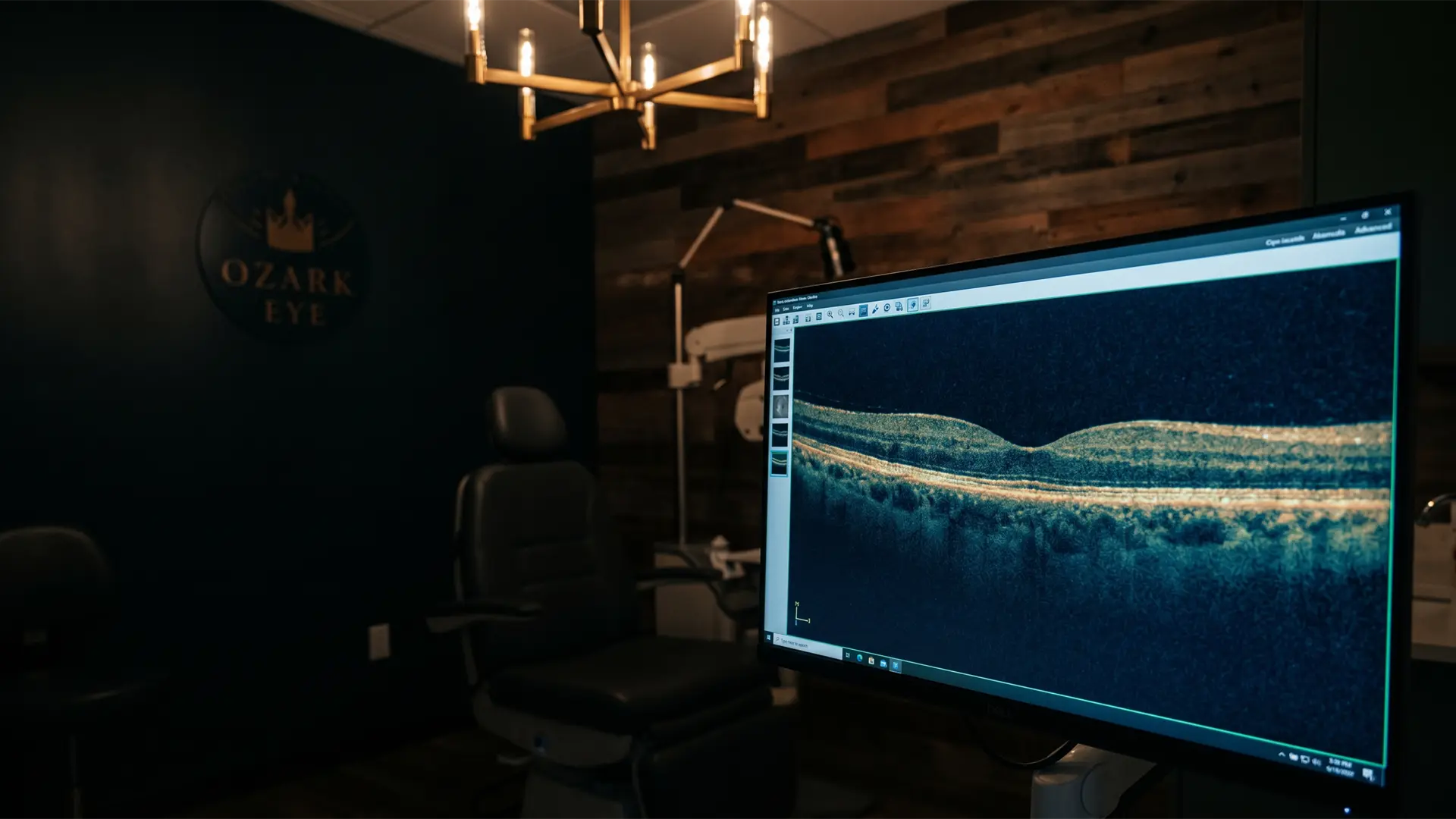
A non-invasive scan that produces high-resolution cross-section images of the retina and optic nerve — similar to an MRI for the eye, without radiation. OCT detects glaucoma damage, macular degeneration, diabetic macular edema, and retinal layers in micron-level detail. It's the single most important diagnostic tool for chronic eye disease.
Visual field testing
Measures peripheral vision to map blind spots and detect functional vision loss. Critical for monitoring glaucoma progression, ruling out neurological conditions affecting vision, and detecting damage from medications like Plaquenil. Painless, non-invasive, takes about 5 minutes per eye.
Digital retinal imaging
Wide-field photographs of the entire retina, documented and stored over time. We compare year-over-year images to catch subtle changes — new blood vessels, hemorrhages, drusen, lesions — before they affect vision. Often replaces or supplements dilation.
Tonometry & pachymetry
Tonometry measures intraocular pressure (the main glaucoma risk factor). Pachymetry measures corneal thickness — a critical adjustment factor for accurate pressure readings, and an independent risk factor for glaucoma. Both are part of every comprehensive exam.
Corneal topography
Maps the exact shape and curvature of the cornea. Essential for specialty contact lens fitting (scleral, custom RGP, ortho-K), detecting keratoconus and irregular corneal conditions, and planning for cataract or refractive surgery. See specialty contact lenses →
Slit-lamp biomicroscopy
The workhorse of every eye exam — a high-magnification microscope with adjustable lighting that lets us examine the front and back of the eye in detail. Used for diagnosing dry eye, infections, inflammation, foreign bodies, cataracts, and dozens of other conditions.
Every comprehensive exam includes the diagnostics appropriate to your age, history, and risk factors. We don't run unnecessary tests — but we do run the ones that catch disease early.
Your eye disease care journey.
Whether you're being screened for the first time or transferring care for an existing diagnosis, here's how chronic eye disease management works at Ozark Eye.
Baseline evaluation
Comprehensive exam with dilated retinal evaluation, OCT imaging, intraocular pressure check, and any condition-specific testing (visual fields for glaucoma, macular OCT for AMD, etc.). If you're transferring care, bring records from your prior provider — we'll incorporate them.
Diagnosis & treatment plan
Dr. Daiber reviews findings with you in plain language. If treatment is needed — prescription drops, monitoring schedule, lifestyle changes, or referral for surgical care — you'll leave with a clear written plan.
Ongoing monitoring
Most chronic conditions are monitored every 3 to 12 months depending on severity. We track changes over time with comparison imaging and adjust treatment as needed.
Specialist co-management
When surgery or advanced intervention is needed (cataract surgery, anti-VEGF injections for wet AMD, glaucoma laser or MIGS), we refer to trusted local specialists and continue your routine care between specialist visits — so you don't have to drive an hour for every follow-up.
Common questions about eye disease management and treatment
Is eye disease care billed to vision or medical insurance?+
Chronic eye conditions — glaucoma, macular degeneration, diabetic eye disease, cataracts, eye infections — are medical, not vision care. We bill your medical insurance (BCBS, Medicare, Aetna, Humana, etc.) for these visits, not your vision plan (VSP, EyeMed, etc.). Most medical plans cover these visits with a regular copay. We verify benefits before your visit.
Can an optometrist treat eye disease, or do I need an ophthalmologist?+
Optometrists diagnose and medically manage most eye diseases — including prescribing medications, monitoring with advanced imaging, and adjusting treatment over time. Surgical interventions (cataract surgery, glaucoma surgery, vitrectomy) require an ophthalmologist, but the diagnosis, ongoing monitoring, pre-op planning, and post-op care all happen at your optometrist. For most chronic conditions, your optometrist is your primary eye care provider and an ophthalmologist becomes involved only if surgery is needed.
Do I need a referral to be seen for an eye disease?+
No referral needed in most cases — patients can self-refer to Ozark Eye for any eye-related concern. If your specific medical insurance plan requires a referral, our staff will help you sort it out before the visit. Just give us a call.
How often should I be monitored if I have eye disease?+
Schedule depends on condition and severity. Glaucoma: every 3 to 6 months. Macular degeneration: every 6 to 12 months for dry AMD; more frequent if there are signs of progression. Diabetic retinopathy: at least annually for all diabetics; every 3 to 6 months if retinopathy is present. Cataracts: annual until surgery is considered. Dr. Daiber sets your monitoring schedule based on your specific situation.
What is OCT imaging and why does it matter for eye disease?+
OCT (optical coherence tomography) is a non-invasive scan that produces high-resolution cross-section images of the retina and optic nerve — similar to an MRI but for the eye, and without radiation. OCT detects glaucoma damage, macular degeneration changes, diabetic macular edema, and other retinal conditions years before they would be visible to the human eye or affect your vision. It's the single most important diagnostic tool for chronic eye disease, and we use it routinely.
Can eye exams detect other health problems?+
Yes — frequently. The retina is the only place in the body where blood vessels and nerves can be observed directly. We routinely catch early signs of diabetes, high blood pressure, high cholesterol, and occasionally autoimmune conditions or even brain tumors. For patients without a primary care physician, we coordinate findings and recommend follow-up where appropriate.
I was diagnosed with eye disease elsewhere. Can I transfer care to Ozark Eye?+
Absolutely. We accept transfer patients with existing eye disease diagnoses and welcome second opinions. Bring records from your prior provider (or have them faxed) and we'll incorporate them into your baseline evaluation. Continuity of care matters, especially for chronic conditions.
What if I need surgery?+
For surgical eye care (cataract surgery, glaucoma surgery, retinal procedures), Dr. Daiber refers you to a trusted local surgical specialist and continues your pre- and post-operative care right here in Pea Ridge. You get convenient follow-up close to home, and one familiar doctor through the entire process — fewer drives to Fayetteville or Rogers for routine checks.
Concerned about eye disease? Get evaluated.
If you have risk factors, a family history, or symptoms that concern you — book a comprehensive evaluation. Early detection is the difference between preventable and permanent.
Let's see what good vision can do.
Book online in under a minute, or call us directly. New patients welcome.